Patterns of Lung Disease
WHAT ARE THE PATTERNS OF RESPIRATORY DISEASE?
Doctors often speak of many lung diseases as representing
one of two patterns of breathing: restrictive or obstructive.
These terms refer only to how a respiratory problem affects a
patient's breathing pattern; they say nothing about cause, treatment,
Xray appearance, or prognosis of his condition. Furthermore,
the two breathing patterns frequently are seen together in one
disease. At best they are merely descriptive of many respiratory
problems. Despite their limitations, use of the terms "restrictive"
and "obstructive" is so pervasive that they will be
defined here and used throughout this book.
WHAT IS RESTRICTIVE RESPIRATORY DISEASE?
This is any respiratory condition where the patient is unable to take in a full, deep breath. It can be due to lung, chest cage, or nervous system disease. By analogy imagine a steel hoop placed around your chest so that you can breathe in a little, but not take a full, deep breath. Your breathing is then restricted. The closer the steel hoop comes to your resting breathing level at the end of a normal breath, the less you can inspire and the more severe is the restriction.
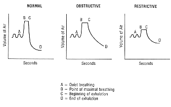
Any respiratory condition resulting in inability to expand fully the lungs is a restrictive problem. Once air is inhaled, however, patients with restrictive disorders can exhale without any impediment or obstruction. Hence, the major distinction between restrictive and obstructive lung disease is between difficulty getting all the air in (restrictive) and getting all the air out (obstructive), This difference can be appreciated by measuring the forced vital capacity (Figure 1).
WHAT IS OBSTRUCTIVE LUNG DISEASE?
Asthma, chronic bronchitis, and emphysema are examples of obstructive lung disease. Characteristic of this group is difficulty getting all the air out. As a group obstructive lung diseases are the greatest cause of respiratory morbidity in the United States. Obstructive lung disease is best diagnosed by a simple pulmonary function test of the forced vital capacity (see Section I). As shown in Figure 1, the obstructed patient can take a deep breath but the rate of exhalation is slowed. This is contrasted with a restricted patient, who cannot inhale as much air but can exhale it readily. For comparison the normal FVC curve is also shown.
Figure 1. Patterns of respiratory disease as shown
by measurement of forced vital capacity.

Respiratory diseases and conditions commonly associated
with a restrictive breathing pattern are listed in Table 1. Although
many of the lung diseases listed here may also be associated with
airway obstruction, restriction is usually predominant.
| ||||||||||||||||||||||||||||
| ||||||||||||||||||||||||
WHAT IS THE DIFFERENCE BETWEEN UPPER AND LOWER AIRWAY OBSTRUCTION?
Asthma, chronic bronchitis, and emphysema are all diseases of the lower airways (bronchial tubes). The trachea, larynx (voice box), and pharynx (area behind the tongue) comprise the upper airway and are not involved in these conditions. Croup,laryngitis, tracheitis, and epiglottitis are infections or inflammations of the upper airway; they may lead to upper airway obstruction. They are more common in children than in adults; also, children are more prone to upper airway obstruction because of the child's smaller airway.
A tumor or foreign body can also obstruct the upper airway. Any patient with upper airway obstruction can present with expiratory wheezing just like asthma, so the distinction is important, Accurate diagnosis of upper airway obstruction can usually be made by a combination of history and physical examination, Xrays of the neck, and spirometry. Stridor (a highpitched, inspiratory wheeze) is common in upper airway obstruction and is not usually heard with asthma and other forms of lower airway obstruction. (Complete airway obstruction is a medical emergency).
Note that central nervous system and chest wall conditions
do not ordinarily lead to an obstructive breathing pattern, whereas
they are a common cause of a restrictive pattern. This is because the
process of expiration is passive and requires only normal airways
once the air has been inhaled. By contrast, the process of inspiration
is active, requiring an intact brain control center, chest wall,
and lungs. Finally, some diseases, such as sarcoidosis (Section O),
can manifest both an obstructive and restrictive pattern.
[Return to Table of Contents]